
Wednesday, October 7, 2015
Readers Choice Award
We are excited to announce that our orthopedic group has been chosen for the Gazette Readers Choice award. Congratulations and thank you to all of the staff and doctors that have helped us take care of so many people. Keep up the great work!
Monday, July 20, 2015
Coaches can be a strong influence in preventing football injuries, say researchers | EurekAlert! Science News
Coaches can be a strong influence in preventing football injuries, say researchers | EurekAlert! Science News

This is exactly why programs such as the Northampton Cal Ripken League are so important. Programs that coach the Coaches are critical for youth sports and vital for our kids.
Friday, July 10, 2015
Achilles Tendon Injuries
Achilles tendon and calf muscle tears are problems that have recently become near and dear to my heart. This, much like tennis elbow is the source of many athletic midlife crises. The Achilles tendon is actually a combination of three separate tendons. The Gastrocnemius is the big, bulbous muscle that gives the calf muscle shape. The Soleus is a flat muscle that sort of resembles a fillet of sole, and the Plantaris is a very small muscle that does not seem to provide any substantial function. The tendons of these three muscles come together to form the thick heel cord that is the source of many problems.

Unfortunately, as we age the healing process is not quite enough to support the constant pounding that the Achilles tendon receives. When an injury occurs, it is typically described as a feeling of being kicked in the back of the leg. Typically these tendons are injured during an explosive events such as jumping, landing or starting a "sprint". The typical age for this type of injury is between 40 and 50 years old and occurs predominantly in male patients. Up until recently, the recommendation was almost universally for surgical repair. The rationale behind this was to decrease the rate of rerupture. Now, with more advanced rehabilitation techniques, we are able to spare many people the pain of surgery. In the highly athletic population however, there is still the belief that surgical repair improves the strength and function in the long run. The recovery from any type of Achilles tendon rupture is quite long. It frequently takes 6-12 months to completely recover from such an injury and requires intensive rehab.
-J Fallon
-J Fallon
Tuesday, June 2, 2015
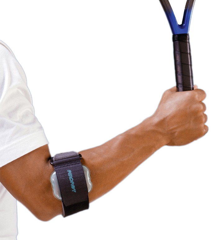
But I Don't Even Play Tennis!
Tennis elbow is a frustratingly annoying problem that is life's little "Welcome to Mid-Life!" gift. It has been referred to as some derivation of tennis elbow since the 1880's. The name came about not because it was believed to be caused by playing tennis, but because the physician that published essays in the 1880's about this problem noted that playing tennis seemed to make it more painful. Since then, the moniker has stuck. The true cause is usually quite variable: tapping sugar maples, crochet, working power lines... it rarely is directly caused by tennis. Ultimately, the problem is that you no longer heal faster than you are injuring yourself. What occurs is that the tendons that attach the extensor muscles of your wrist to the lateral part of your elbow sustain "Microtrauma" through your daily over-use (tapping maples trees as an example). In the ideal 18 year old elbow, this microtrauma would heal perfectly, without you noticing. Unfortunately, as we age, we are unable to heal as effectively and instead of healing with normal tendon, we form a less organized structure that vaguely resembles tendon. These microtrauma events pile up and eventually you end up with lateral epicondylitis, traditionally referred to as Tennis Elbow. There are literally hundreds of "treatments" for lateral epicondylitis. Like many other things in medicine (the common cold) if there are a bunch of remedies, nothing works for everyone.
Monday, May 11, 2015
Do I need an MRI before I see an Orthopedic Surgeon?
The short answer to this is no. Well, not usually. MRIs are a very useful tool in orthopedic surgery, and we have come to depend on them for many facets of diagnosis and surgical planning.

There is commonly the thought that obtaining an MRI before your visit will "Save a step" in getting you better. What I frequently tell patients is that "I treat people, not MRIs". What that means is that I am far more interested in what you have to say and what your knee feels like, than what an MRI can tell me about your knee. So, getting an MRI may be necessary at some point, but may not be necessary.

MRIs also come in different varieties and I order special MRI sequences depending on the question I want answered. For example, if I suspect a SLAP tear in a young pitcher I will order dye injected into the shoulder joint prior to the MRI, making it an MR arthrogram. I will also request that their arm be placed in a certain position. I will not typically ask for an MR arthrogam in a 50 year old carpenter with shoulder pain in which I suspect a rotator cuff tear. If a patient has had previous surgery, metallic anchors would make an MRI useless because the metal would interfere with the magnets and the picture would be blurry. In that case, a CAT scan with dye injected into the shoulder would be a more useful way to see the structure of the shoulder.
The final reason not to try to get an MRI before you see me is that most primary care doctors have difficulty convincing your insurance that they should pay for an MRI. Their staff has to argue and struggle with the insurance companies to get the MRI "pre-approved". Frequently, despite all of their efforts, the insurance companies feel that the MRI is not warranted. Our office, on the other hand has a near flawless record of getting these approved. Part of this is my wonderful staff and their familiarity with the nuances of MRI scheduling and part of it is that for better or worse, my recommendation as a specialist carries more weight with the insurance companies than your PCP. So, let us do the work for you and save yourself the aggravation.
- J. Fallon
There is commonly the thought that obtaining an MRI before your visit will "Save a step" in getting you better. What I frequently tell patients is that "I treat people, not MRIs". What that means is that I am far more interested in what you have to say and what your knee feels like, than what an MRI can tell me about your knee. So, getting an MRI may be necessary at some point, but may not be necessary.
MRIs also come in different varieties and I order special MRI sequences depending on the question I want answered. For example, if I suspect a SLAP tear in a young pitcher I will order dye injected into the shoulder joint prior to the MRI, making it an MR arthrogram. I will also request that their arm be placed in a certain position. I will not typically ask for an MR arthrogam in a 50 year old carpenter with shoulder pain in which I suspect a rotator cuff tear. If a patient has had previous surgery, metallic anchors would make an MRI useless because the metal would interfere with the magnets and the picture would be blurry. In that case, a CAT scan with dye injected into the shoulder would be a more useful way to see the structure of the shoulder.
The final reason not to try to get an MRI before you see me is that most primary care doctors have difficulty convincing your insurance that they should pay for an MRI. Their staff has to argue and struggle with the insurance companies to get the MRI "pre-approved". Frequently, despite all of their efforts, the insurance companies feel that the MRI is not warranted. Our office, on the other hand has a near flawless record of getting these approved. Part of this is my wonderful staff and their familiarity with the nuances of MRI scheduling and part of it is that for better or worse, my recommendation as a specialist carries more weight with the insurance companies than your PCP. So, let us do the work for you and save yourself the aggravation.
- J. Fallon
Sunday, May 10, 2015
What Kind of Music Do You Play in the Operating Room?
I was recently asked this by one of my high-school aged patients. My response was, "It depends... what do you want to listen to while I operate on you?". So we spent the next 45 minutes exploring the K-Pop station on Pandora.

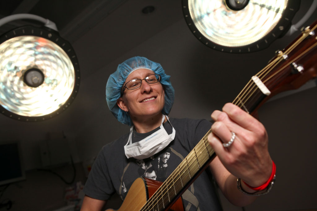
Some days require something mellow like Jack Johnson or Big Head Todd. Other days need a kick in the pants and we get Ja Rule. Most days end with AC/DC and you don’t want to hear my OR Jazz mix… that’s a bad day. Music is good for me, it is good for my team and it also good for the patient. Playing melodies for patients has been shown to decrease anxiety levels and heart rates in perioperative patients.
Fun fact: Apollo was the god of Music and Healing
Monday, May 4, 2015
The Biology of ACL Reconstruction and Regeneration
One of the best examples of the wonders of modern medicine is Anterior Cruciate Ligament reconstruction. Because the body is unable to heal the ACL on its own, it requires reconstruction in order to restore normal function to the knee. The process of ACL regeneration relies heavily on the biology of your knee.
 When I reconstruct your ACL, I take a tendon, either from an organ donor or from a different part of your body, then place it where I want a ligament to grow. Your body uses this tendon as a scaffolding to regenerate a new anterior cruciate ligament. This process occurs and 3 phases. The initial phase is characterized by necrosis or cell death. During the first 4 weeks after surgery, your body tears down all of the living cells in the graft, leaving only nonliving tissue as a blueprint for the final ligament. The second portion of the regeneration process is called the proliferation phase. This usually occurs in the second and third month after surgery. This is characterized by influx in your own body’s cells into the remaining scaffolding. The scaffolding also undergoes changes that cause significant weakness in the graft. 6-8 weeks after surgery is typically thought of as the weakest point and the time where of the structure of the graft needs the most protection. The final phase, the ligamentization phase begins about 3 months after surgery. During this time, the graft is slowly and steadily getting stronger and becoming more like the original ACL. There is no clear end point however there is plenty of evidence suggesting that the graft will continue to mature over the following year.
Subscribe to:
Posts (Atom)